Urinary bladder is a muscular reservoir of urine, which lies in the anterior part of the pelvic cavity. It varies in in its size, shape and position according to the amount of urine it contains.
EXTERNAL FEATURES OF URINARY BLADDER
Empty bladder has following features :-
- Apex – directed forwards
- Base – directed backwards
- Neck – lowest and fixed
- Three surfaces – superior, right and left inferolateral
- Four borders – two lateral, one anterior and one posterior
Full bladder has following features :-
- Apex – directed upwards
- Neck – directed downwards
- Two surfaces – anterior and posterior
LIGAMENTS OF THE URINARY BLADDER
True ligaments are :-
- Lateral true ligament – It extends from side of bladder to the tendinous arch of the pelvic fascia.
- Lateral puboprostatic ligament – It extends from anterior end of the tendinous arch of the pelvic fascia to the upper part of the prostatic sheath.
- Medial puboprostatic ligament – It extends from back of the pubic bone to the prostatic sheath.
- Medial umbilical ligament – It is the remnant of the urachus.
- Posterior ligament – It extends from each side of bladder to wall of pelvis.
False ligaments are :-
- Median umbilical fold
- Medial umbilical fold
- Lateral false ligament
- Posterior false ligament
INTERIOR OF THE URINARY BLADDER
Examined by – Cystoscopy, at operation or at autopsy

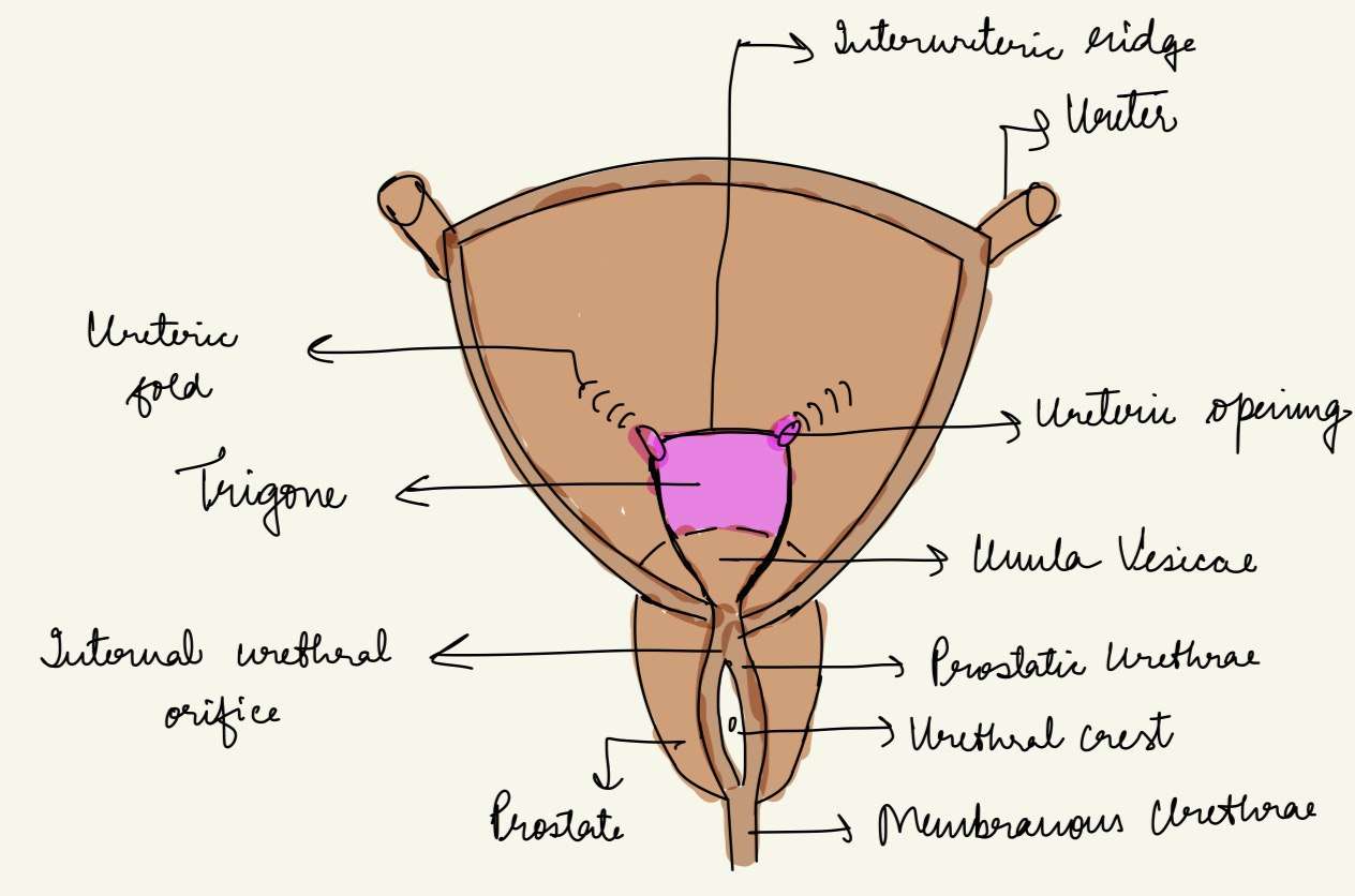
Trigone of the bladder
Small triangular smooth area over the lower part of the base of bladder is known as the trigone of bladder. The apex of the trigone is directed downwards and forwards. The internal urethral orifice, opening into the urethra, is located here.
Uvula vesicae – It is the slight elevation on the trigone immediately posterior to the urethral orifice, produced by the median lobe of the prostate.
Bar of mercier– The base of the trigone is formed by the interureteric ridge or bar of mercier produced by continuation of the inner longitudinal muscle coats of the two ureters.
CAPACITY OF THE BLADDER
- Mean capacity if the bladder in an adult male is 220 ml (120-320 ml).
- Filling beyond 220 ml causes the desire to micturate.
- Filling beyond 500 ml causes referred pain in the lower part of the abdominal wall, perineum and penis.
ARTERIAL SUPPLY
Main supply – Superior and Inferior vesical arteries, branches of anterior trunk of the internal iliac artery. Additional supply – In males, from the obturator and inferior gluteal arteries and in females, from the uterine and vaginal arteries.
VENOUS DRAINAGE
There is a vesical venous plexus lying on the inferolateral surfaces of the bladder which drains into the internal iliac veins.
LYMPHATIC DRAINAGE
Most of the lymphatics from the urinary bladder terminate in the external iliac nodes. A few vessels may pass to the internal iliac nodes or to the lateral aortic nodes.
NERVE SUPPLY
The urinary bladder is supplied by vesical plexus of nerves. The vesical plexus contains both sympathetic and parasympathetic, each of which contains motor and sensory fibres.
- Parasympathetic efferent fibres (S2-S4) are motor to the detrusor muscle. If these are destroyed, normal micturition is not possible.
- Sympathetic efferent fibres (T11-L2) are said to be inhibitory to the detrusor.
- The somatic pudendal nerve supplies the sphincter urethrae which is voluntary and situated within the wall of urethra
- Sensory nerves: Pain sensation caused by distention or spasm of the bladder wall are mainly carried by parasympathetic nerve and partly by sympathetic nerve.
CLINICAL ANATOMY OF URINARY BLADDER
- Cystoscope – For the examination of the interior of the bladder.
- The distended bladder may be ruptured by injuries of the lower abdominal wall.
- Hypertrophy of bladder – It is caused due to chronic obstruction to the outflow of urine by an enlarged prostate.
- Suprapubic cystotomy – The bladder is distended with about 300 ml of fluid. As a result, the anterior aspect of the bladder comes in direct contact with the anterior abdominal wall, and can be approached without entering the peritoneal cavity.
- Emptying of bladder – It is essentially a reflex function. acute injury to the cervical/thoracic segments of spinal cord leads to a state of spinal shock. The muscle of the bladder and the sphincter urethrae is relaxed but sphincter urethrae is contracted. The bladder distends and the urine dribbles. Damage to the sacral segments of spinal cord situated in lower thoracic and lumbar one vertebra results in autonomous bladder. Continuous dribbling occurs.
- Urinary bladder is one of the sites for stone formation as concentrated lies here.
FOR ANATOMY SECTION VISIT – https://medmaps.in/category/notes/anatomy/
GET CONNECTED TO US ON OUR INSTAGRAM PAGE – https://www.instagram.com/medmaps.in/


0 Comments