INTRODUCTION
Foetal circulation is equally as essential as the onset of breathing at birth are immediate circulatory adjustments that allow adequate blood flow through the lungs.
Also, circulatory adjustments during the first few hours of life cause more and more blood flow through the baby’s liver, which up to this point has had little blood flow.
ANATOMICAL STRUCTURE OF THE FETAL CIRCULATION
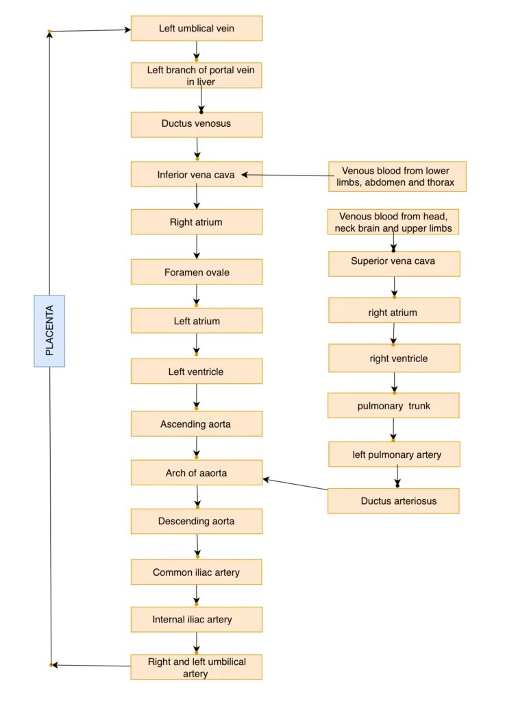
•During fetal life the lungs are mainly non-functional and liver is only partially functional.
•So it is not necessary for the fetal heart to pump much blood through either the lungs or the liver.
•However, the fetal heart must pump large quantities of blood through the placenta.
•Therefore, special anatomical arrangements cause the fetal circulatory system to operate much differently from that of the newborn baby.
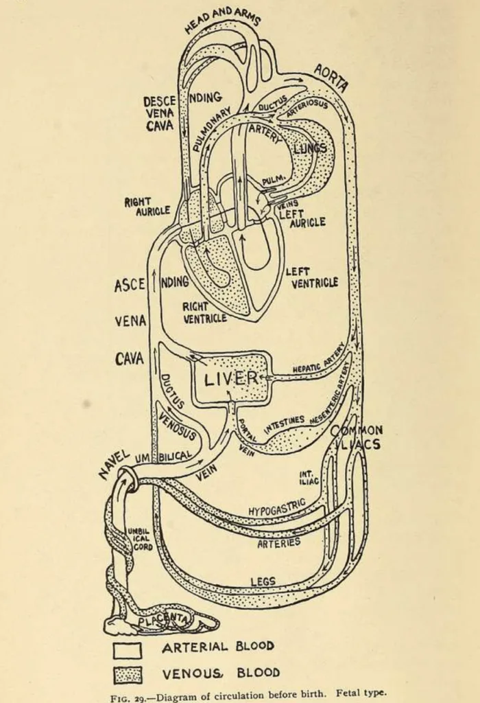
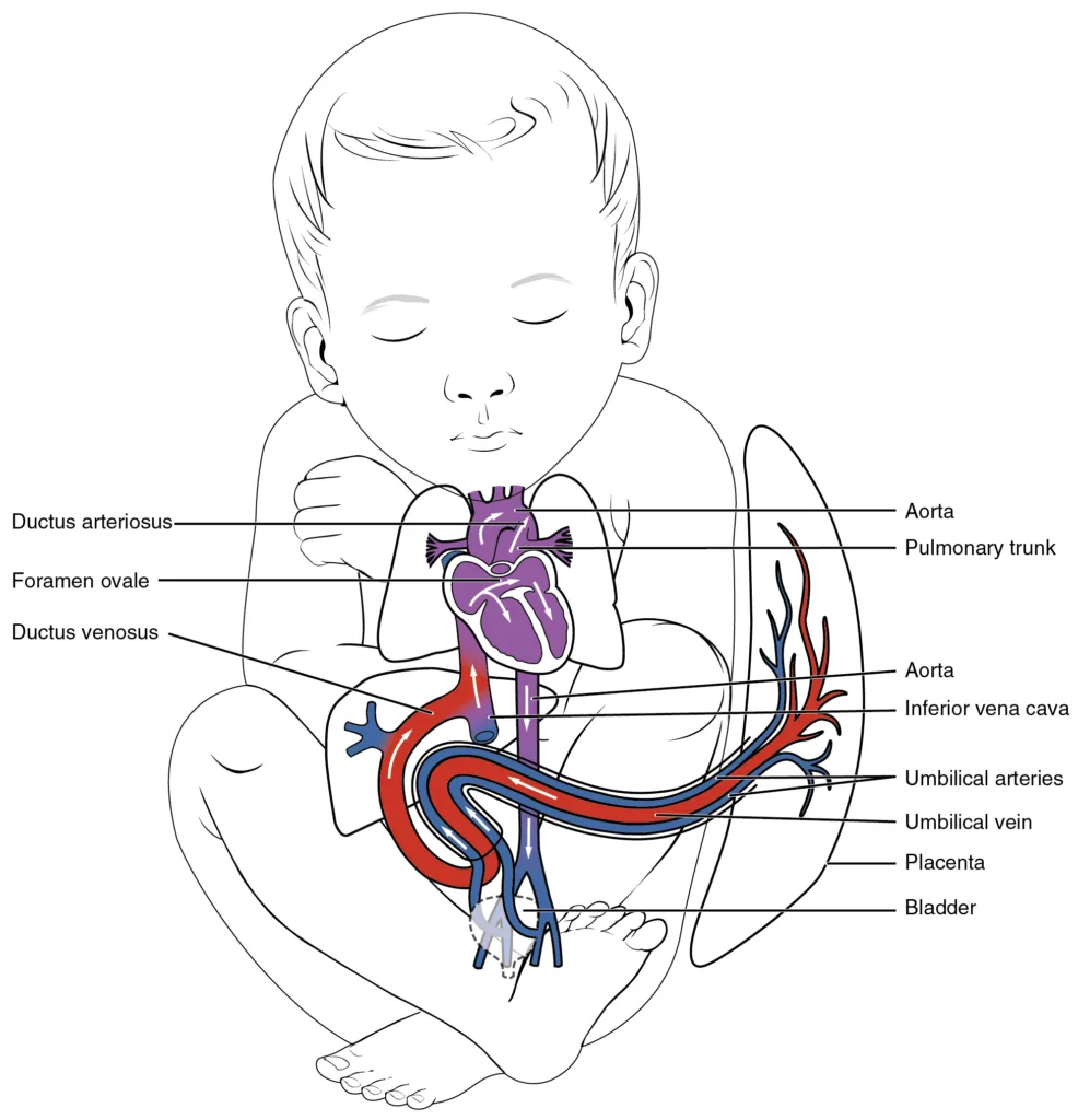
•Blood returning from the placenta through the umbilical vein passes through the ductus venosus, thus bypassing mainly liver.
•Then most of the blood entering the right atrium from inferior vena cava is directed in a straight pathway across the posterior aspect of the right atrium and through the foramen ovale directly into the left atrium.
•Thus, the well- oxygenated blood from the placenta enters mainly the left side of the heart, rather than the right side of the heart.
•And is pumped by the left ventricle mainly into the arteries of the head and forelimbs.
•The blood entering the right atrium from the superior vana cava is directed downward through the tricuspid valve into the right ventricle.
•This blood is mainly deoxygenated blood from the head region of the fetus.
•It is pumped by the right ventricle into the pulmonary artery.
•Then mainly through the ductus arteriosus into the descending aorta.
•Then through the two umbilical arteries into the placenta, where the deoxygenated blood becomes oxygenated.
CHANGES AT BIRTH
1.Decreased pulmonary and increased systemic vascular resistance at birth.
(i)The primary changes in the circulation at birth are, first, loss of the tremendous blood flow through the placenta, which approximately doubles the systemic vascular resistance at birth.
This increases the aortic pressure, as well as the pressure in the left ventricle and atrium.
(ii)Second, the pulmonary vascular resistance greatly decreases as the result of expansion of the lungs.
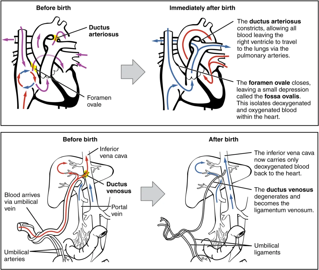
2. Lungs start functioning.
3. Umbilical vein forms ligamentum teres.
4. Ductus venosus forms ligamentum venosum.
5. Foramen ovale closes.
6. Ductus arteriosus form ligamentum arteriosum.
7.Umbilical arteries form medial umbilical ligament.
8. Placenta is delivered and removed.
FOR ANATOMY SECTION VISIT – https://medmaps.in/category/notes/anatomy/
GET CONNECTED TO US ON OUR INSTAGRAM PAGE – https://www.instagram.com/medmaps.in/


0 Comments