Internal capsule is a large band of fibres which serves as a vital conduit for communication between different regions of the central nervous system. Its intricate pathways facilitate the transmission of motor, sensory, and cognitive signals, playing a pivotal role in orchestrating our every movement, perception, and thought.
ANATOMY OF INTERNAL CAPSULE
Location
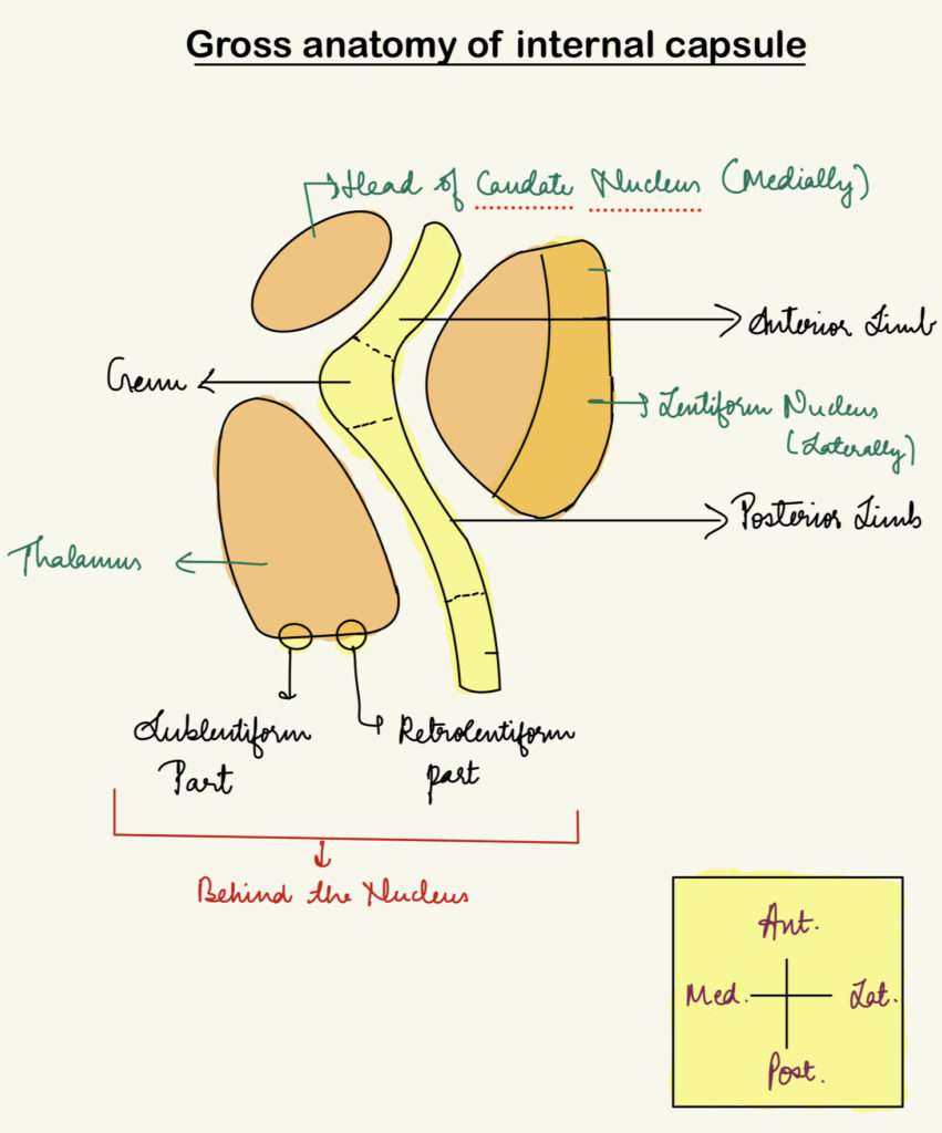
Situated in the inferomedial part of each cerebral hemisphere. In the horizontal cross-section of the brain, it appears V-shaped with its concavity directed laterally. The concavity is occupied by the lentiform nucleus.
Subdivisions of internal capsule

The internal capsule is divided into the following parts :
- Anterior Limb – It lies between the head of the caudate nucleus and the lentiform nucleus.
- Genu – It is the band between the anterior and posterior limbs.
- Posterior Limb – It lies between the thalamus and lentiform nucleus.
- Sublentiform part – It lies below the lentiform nucleus.
- Retrolentiform part – It lies behind the lentiform nucleus.
Note : Sublentiform part can be seen in a coronal section, whereas the rest of the parts are seen in a horizontal section of brain.
Boundaries and surrounding structures
- Anteriorly : Anterior limb is bounded by the head of caudate nucleus.
- Posteriorly: The posterior limb of the internal capsule is bordered by the thalamus and the lenticular nucleus (which includes the putamen laterally and the globus pallidus medially).
- Superiorly: It extends up to the lateral ventricle and the corona radiata.
- Inferiorly: The internal capsule descends toward the cerebral peduncle in the brainstem.
The surrounding of internal capsule is formed by the thalamus, basal ganglia (laterally), lateral ventricles (superiorly) and corona radiata.
FIBRES OF INTERNAL CAPSULE
Motor fibres
- Corticopontine fibres – It lies in anterior limb, genu and posterior limb.
- Frontopontine fibres – It is a part of corticopontocerebellar pathway. They start from the frontal lobe and reach the opposite cerebellar hemisphere through pontine nuclei.
- Parietopontine and occipitopontine fibres – They lie in retrolentiform part of internal capsule.
- Temporopontine fibres – It lies in sublentiform part of internal capsule.
Pyramidal fibres
- Corticonuclear fibres – Corticonuclear fibers project to the motor nuclei of the cranial nerves located in the brainstem. These motor nuclei include motor nuclei of cranial nerve V, VII, IX, X, XI, XII.
- Corticospinal fibres – Fibres for anterior horn cells of muscles of head and neck lie in genu.
Extrapyramidal fibres
- Corticostriate fibres
- Corticorubral fibres
These fibres start from cerebral cortex and reach corpus striatum and red nucleus.
Sensory fibres
- Anterior thalamic radiation – Fibres from anterior and dorsomedial nuclei of thalamus terminate in cortex of the frontal lobe.
- Superior thalamic radiation – Fibres of ventral group of nuclei of thalamus, reach sensory areas of frontal and parietal lobes.
- Inferior thalamic radiation – It connects medial geniculate body with primary auditory cortex.
- Posterior thalamic radiation – These fibres connect lateral geniculate body to area 17 forming optic radiation.
BLOOD SUPPLY OF INTERNAL CAPSULE

Anterior limb
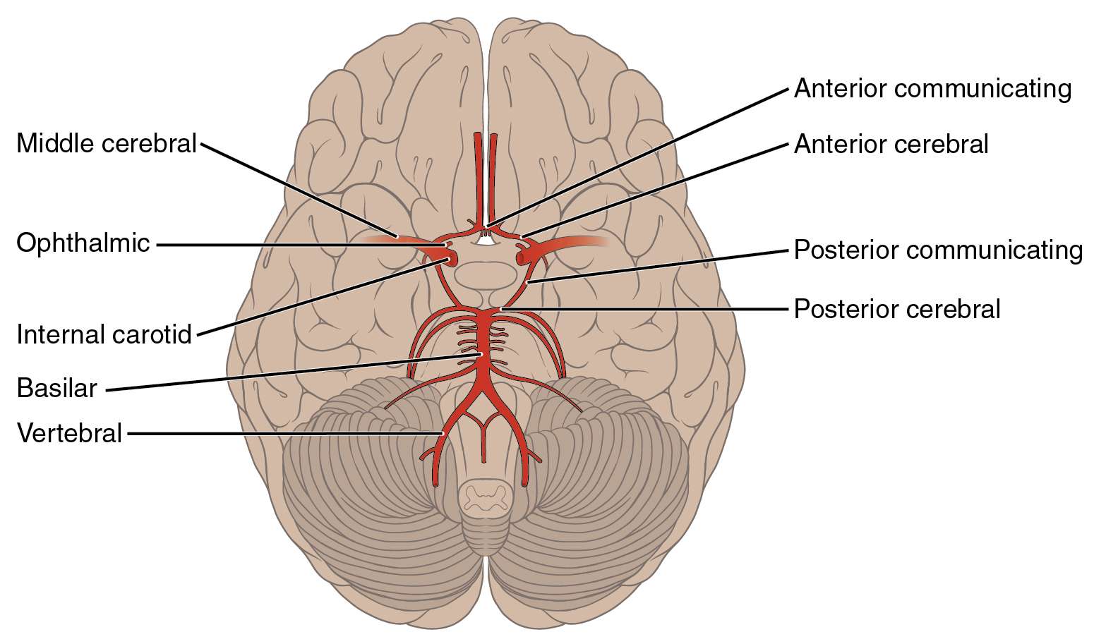
- Recurrent branch of anterior cerebral
- Direct branch from anterior cerebral
Genu
- Direct branch from internal carotid
- Posterior communicating
Posterior limb
- Lateral striate branches of middle cerebral
- Medial sriate branches of middle cerebral
- Branch of anterior choroidal
Sublentiform part
- Branches of posterior cerebral
- Branch of anterior choroidal
Retrolentiform part
- Branch of posterior cerebral
CLINICAL ANATOMY OF INTERNAL CAPSULE
- Thrombosis of recurrecnt branch of anterior cerebral artery – It leads to upper motor neuron type of paralysis of the opposite upper limb and of the fac e.
- Lesion of internal capsule – It is usually vascular, due to involvement of the lateral striate branches of the middle cerebral artery. They give rise to hemiplegia on the opposite side of the body. It is an upper motor neuron type of paralysis. The largest lateral striate artery is called ‘Charcot’s artery of cerebral hemorrhage’.
FOR ANATOMY SECTION VISIT – https://medmaps.in/category/notes/anatomy/
GET CONNECTED TO US ON OUR INSTAGRAM PAGE – https://www.instagram.com/medmaps.in/


0 Comments