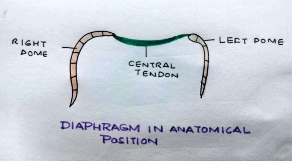
Thoracoabdominal diaphragm is a dome shaped muscle forming the partition between the thoracic and abdominal cavities. It is the chief muscle of respiration. The diaphragm is a crucial muscle that plays a central role in the process of breathing and is located at the base of the thoracic cavity. As the primary muscle of respiration, the diaphragm contracts and relaxes to facilitate the inhalation and exhalation of air.

ORIGIN OF THORACOABDOMINAL DIAPHRAGM
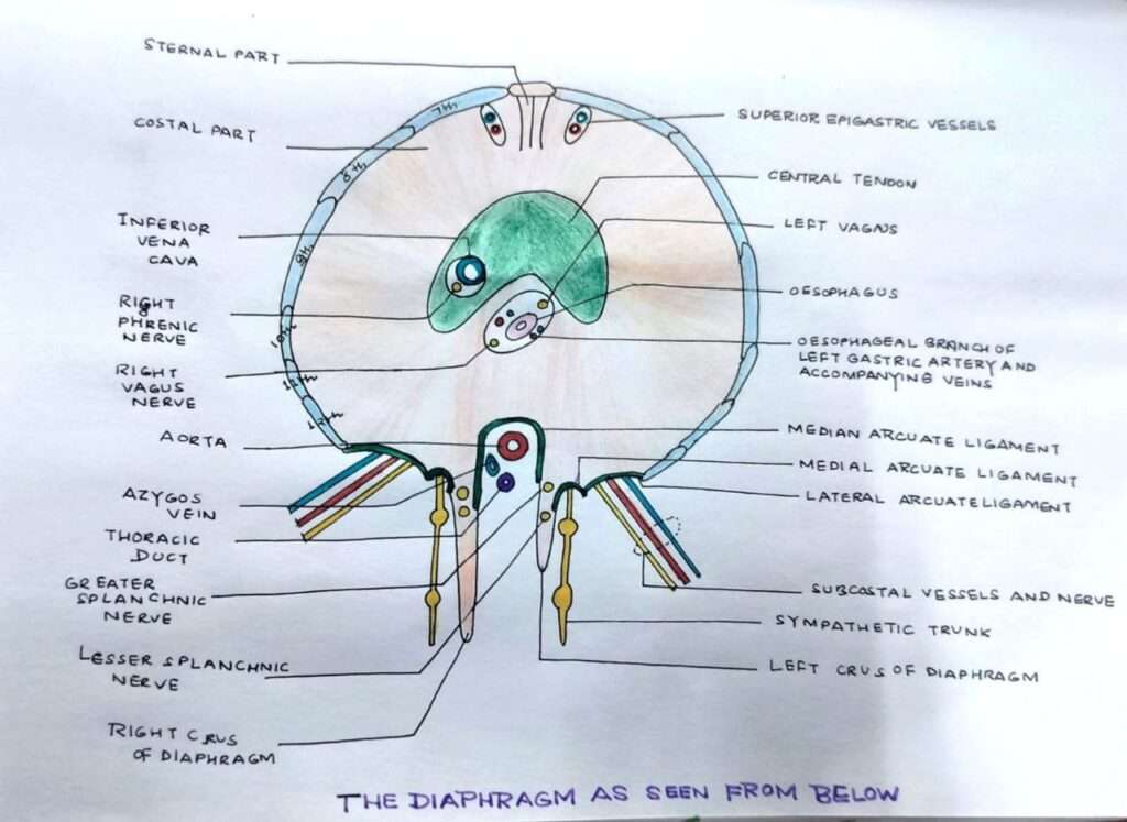
The muscle fibres are grouped into 3 parts :-
- Sternal part – From two fleshy slips from the back of xiphoid process.
- Coastal part – From the lower 6 ribs on each
- Lumbar part – From the medial and lateral lumbocostal arches and from the lumbar vertebrae by right and left crura.
INSERTION OF THORACOABDOMINAL DIAPHRAGM
Insertion of it takes place at central tendon which has the following characteristics :-
- Lies below the pericardium
- Trilobar in shape
- The central area consists of four well marked diagonal bands.
OPENINGS IN THE THORACOABDOMINAL DIAPHRAGM

There are 3 large openings :-
1.Aortic opening – Level – T-12
Transmits – Aorta, thoracic duct and azygous vein
2. Oesophageal opening – Level – T-10
Transmits – Oesophagus, right vagus nerve and oesophageal branch of left gastric artery
3. Vena caval opening – Level – T-8
Transmits – Inferior vena cava and right phrenic nerve
There are 7 small openings :-
- Greater and lesser splanchnic nerve
- Sympathetic chain
- Subcoastal nerve and vessels
- Superior epigastric vessels
- Musculophrenic vessels
- Intercoastal nerve and vessels
- Left phrenic nerve
RELATIONS
Superiorly – Pleurae, lungs and the pericardium
Inferiorly – Peritoneum, liver, fundus of stomach, spleen, kidney and suprarenals
NERVE SUPPLY
Motor – Phrenic nerves are the sole motor nerves to thoracoabdominal diaphragm
Sensory – By the phrenic nerves (central part) and the lower 6 thoracic nerves (peripheral part)
ACTIONS
- It is the principle muscle for inspiration
- It acts in all expulsive acts like sneezing, coughing, laughing, crying, etc.
- Sphincteric action in lower end of the oesophagus.
The position of the thoracoabdominal diaphragm depends upon 3 main factors :-
- Elastic recoil of the lung tissue
- Pressure exerted by the abdominal viscera
- While standing, the muscles in the abdominal wall contract, increasing the intra-abdominal pressure.
Because of these three factors , the level of the thoracoabdominal diaphragm is highest in the supine position, lowest while sitting and intermediate while standing.
CLINICAL ANATOMY OF THORACOABDOMINAL DIAPHRAGM
1. Hiccough or hiccup – Result of spasmodic contraction of the thoracoabdominal diaphragm. It may be :- Peripheral – Due to local irritation of thoracoabdominal diaphragm or its nerve. Central – Due to irritation of the hiccough centre in the medulla.
2. Shoulder tip pain – Irritation of the thoracoabdominal diaphragm may cause referred pain in the shoulder because the phrenic and supraclavicular nerves have same root values (C3,C4,C5)
3. Unilateral paralysis of the thoracoabdominal diaphragm – Due to lesion of the phrenic nerve. The paralysed side moves opposite to the normal side.
4. Eventration – Condition in which thoracoabdominal diaphragm is pushed upwards due to congenital defect in the musculature of its left half.
5. Diaphragmatic hernia – It may be congenital or acquired.
FOR ANATOMY SECTION VISIT – https://medmaps.in/category/notes/anatomy/
GET CONNECTED TO US ON OUR INSTAGRAM PAGE – https://www.instagram.com/medmaps.in/


0 Comments